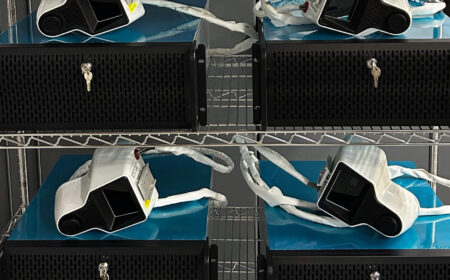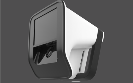
VISIE 3D scanners are Investigational Devices and are not available for sale.

VISIE 3D scanners are Investigational Devices and are not available for sale.
Together, these findings demonstrate the potential of VISIE to transform femoral registration in TKA by providing surgeons with a faster, less invasive, and exposure-independent approach to computer-assisted navigation.
As total knee arthroplasty (TKA) continues to evolve toward more efficient, tissue-sparing procedures, surgeons need navigation technology that adapts to the realities of the patient, not the other way around. Conventional navigation systems often require invasive fixation pins, patient-mounted reference arrays, and manual point-by-point registration, adding surgical steps that can increase procedure complexity and disrupt workflow.
VISIE introduces a pinless registration approach designed to simplify this process while making it less invasive.. Using a rapid, three-dimensional (3D) optical scan, the system registers the exposed distal femoral anatomy to the patient’s preoperative computed tomography (CT) model without the need for fixation pins or manual anatomic registration. The result is a streamlined registration workflow that enables accurate navigation while reducing dependence on extensive anatomic exposure.
To first demonstrate the robustness of the pinless registration approach under intentionally challenging conditions, a porcine knee model was used with only a minimal portion of the distal femur available for optical acquisition. This demonstrated successful registration despite severely restricted anatomic visibility, providing evidence that the scans contain data rich enough to establish anatomic correspondence even from a limited surface.
The pinless registration approach was subsequently demonstrated in a human cadaveric model using a subvastus surgical exposure, which provides only limited visualization of the distal femur. This represents a clinically relevant and demanding registration scenario. Quantitative evaluation under these conditions demonstrated accurate registration despite constrained anatomic exposure, supporting the potential of rapid optical 3D registration for minimally invasive TKA workflows.
Successful navigation begins with accurate registration, yet obtaining a reliable registration can be challenging when only a restricted portion of the femur is visible. Modern tissue-sparing surgical approaches are designed to minimize soft tissue disruption and preserve anatomy, but they also reduce the amount of exposed bone available for registration. As a result, navigation systems must establish an accurate anatomic relationship using limited intraoperative information.
This challenge is further complicated by the differences between the patient’s preoperative CT model and the anatomy visible during surgery. CT-based models represent the underlying bony anatomy, while intraoperative optical scans capture the exposed articular cartilage and surrounding soft tissues. Because these surfaces are not identical, they cannot be directly aligned using conventional surface matching techniques. Differences in cartilage thickness, soft tissue coverage, and anatomic morphology introduce additional complexity that must be addressed to achieve clinically meaningful registration accuracy.
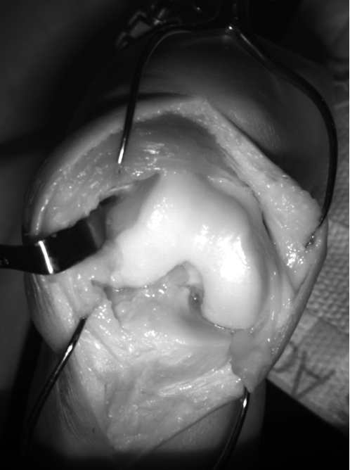
Figure 1. Subvastus knee exposure for procedure. Point of view for the surface scan taken for registration.
The subvastus approach represents one of the most demanding registration scenarios. By preserving the extensor mechanism and minimizing soft tissue disruption, the approach intentionally limits exposure of the distal femur. Much of the anterior and lateral femoral anatomy remains obscured by the vastus medialis, leaving only a small portion of the surface available for optical acquisition. Demonstrating accurate registration under these constrained conditions provides confidence that the technology can perform reliably in the reduced exposures commonly encountered during modern, minimally invasive TKA.
Reliable registration begins with capturing high-quality anatomic data. To support registration in the limited exposures commonly encountered during TKA, VISIE uses a proprietary optical scanning platform designed to maximize the quality and completeness of the acquired surface.
A key feature of the platform is its extended structured light source, which illuminates the surgical field from multiple angles simultaneously. Unlike conventional, lens-based projectors that behave as a single point source of light, the extended illumination geometry improves coverage of anatomic surfaces that would otherwise receive indirect or no illumination. This results in a higher-quality three-dimensional (3D) optical scan with improved signal-to-noise ratio across the exposed anatomy.
Figure 2 highlights the contribution of the extended light source. The regions shown in green and red represent surface points with enhanced signal quality resulting from the multi-angle illumination. By improving the fidelity of the acquired surface data—particularly on anatomically challenging regions—the optical scan provides a stronger foundation for accurate registration, even when only a partial view of the femur is visible.
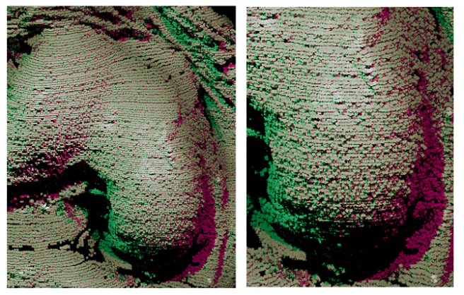
Figure 2. Illustration of the advantage of pattern-projection from VISIE’s unique scanning light source. False-color enhanced points indicate regions of greater signal-to-noise ratio over conventional projection-based 3D scanners, owing to its pattern projection from an extended light source. Scans exhibit greater coverage with lower noise, excellent for anatomic registration to preoperative images.
A key objective of this study was to demonstrate that accurate registration can be achieved even when only a small portion of the distal femur is available for optical acquisition. While typical TKA procedures provide varying degrees of anatomic exposure, challenging cases and tissue-sparing approaches can significantly reduce the visible surface available for registration. To evaluate the limits of the registration method, a porcine knee model was used to create an intentionally demanding registration scenario.
The porcine specimen was prepared so that only a small region of the distal femur remained visible while the majority of the anatomy was obscured (Figure 3, left). This configuration represents a more restrictive field of view than would typically be encountered during standard TKA and serves as a stress test of the registration workflow, to determine whether successful registration could still be achieved.
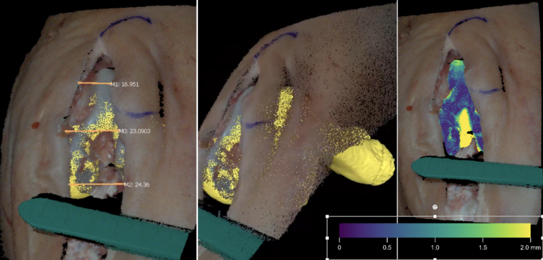
Figure 3. Optical registration of porcine femur to a preoperative CT (yellow) through minimal exposure. Left: Measurements across the incision on the left image are in millimeters. Center: A side view of the registration. Right: False-color indication of distances between the CT scan and the optical scan anatomy after alignment. The scale bar represents the nearest-neighbor-distances between the exposed anatomy in the VISIE scan and the preoperative CT after alignment (right image only).
Despite the limited visible anatomy, the system successfully aligned the intraoperative optical scan with the corresponding preoperative CT model (Figure 3, middle). This alignment remained stable using only the exposed anatomic features, demonstrating that the optical scan information was sufficient to establish an accurate anatomic relationship from limited surface information. Representative images illustrating the available anatomy and resulting registration are shown in Figure 3.
Although this evaluation was not designed to establish registration accuracy relative to an independent ground truth, the quality of the resulting alignment is reflected by a root mean square error (RMSE) of 0.67 mm between the registered optical surface and the corresponding CT-derived anatomy. A representative example of this alignment, including the corresponding surface residuals, is shown in false color in the right panel of Figure 3. This low residual error indicates a close agreement between the aligned surfaces and demonstrates that successful registration can be achieved with the optical scan, despite the intentionally reduced anatomic exposure. These findings provide evidence of the robustness of the registration approach when only a small portion of the distal femur is available for optical acquisition.
Evaluation in a Subvastus Surgical Approach
To evaluate the pre-clinical performance of the registration method, the workflow was assessed using a cadaveric specimen prepared with a subvastus surgical approach. This approach was intentionally selected because it represents one of the most challenging exposure conditions encountered during total knee arthroplasty (TKA), providing only limited visualization of the distal femur while preserving the extensor mechanism (Figure 4).
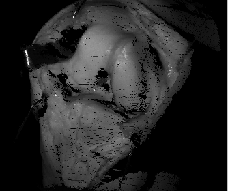
Figure 4. Three-dimensional point cloud of right knee exposure prior to fiducial insertion
A patient-specific CT-based model of the femoral surface was generated and registered with a proprietary algorithm to a high-density optical scan of the exposed anatomy acquired using the VISIE scanning platform. The optical acquisition was completed in less than one second, replacing the traditional point-by-point registration workflow with a rapid anatomic surface capture. Registration was performed using only the exposed anatomic surface, consisting primarily of articular cartilage and adjacent soft tissues. Because this was a cadaveric evaluation, the hip center was simulated for the specimen.
Quantitative Registration Validation
To independently evaluate registration accuracy, the specimen was subsequently resected further to expose additional bony anatomy, and four radiopaque spherical fiducial markers
were implanted in the distal femur (Figure 5). A postoperative CT scan was then acquired, allowing the femur and fiducial locations to be segmented and used as an independent reference model.
The fiducial markers were identified in both the postoperative CT and the optical scan, and the Kabsch-Umeyama algorithm [1] was used to calculate the ground truth transformation between the two coordinate systems. This reference transformation was established with a RMSE of 0.22 mm between corresponding fiducial centroids.
To relate this ground truth back to the original subvastus scan, the pre-fiducial optical scan (Figure 4) was registered to the post-fiducial optical scan (Figure 5). This registration achieved a 91% surface overlap (fitness) with an RMSE of 0.45 mm between the articular surfaces. Combining these transformations established the ground truth CT position within the coordinate system of the original subvastus optical scan, enabling a direct comparison between the anatomic registration and the independently measured reference.
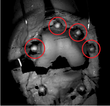
Figure 5. Fiducial placement about the femur. Femoral fiducials circled in red.
Registration Results
Figure 6 illustrates the alignment between the optical scan and the patient-specific CT model following anatomic registration. Compared with the independently established ground truth, the registration demonstrated a rotational difference of 0.70° and a mean translational difference of 1.18 mm at the fiducial locations (Table 1). These results are well within commonly accepted navigation accuracy targets of approximately 2° rotational and 2 mm translational error.
In addition, the VISIE anatomic registration method remains within the accepted navigation accuracy targets for hip center localization errors of up to ±6 mm, a level of accuracy that has been demonstrated to be clinically achievable [3]. This indicates that the observed registration performance is maintained under realistic surgical conditions where hip center estimation is subject to inherent variability. These results are summarized in Table 1.
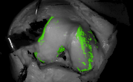
Figure 6. Pre-operative CT (green) registered into the scanner’s coordinate system using VISIE scan (greyscale) and algorithm.
Together, these findings demonstrate that accurate anatomic registration can be achieved despite the limited femoral exposure provided by the subvastus approach. Even with only a portion of the distal femur available for optical acquisition, sufficient anatomic information was captured to establish a precise registration between the intraoperative optical scan and the preoperative CT model. The results also highlight the contribution of the VISIE optical scanning platform. By combining rapid optical acquisition with enhanced illumination of the exposed anatomy, the system provides high-quality surface data that supports accurate registration in exposure-limited surgical workflows, enabling a streamlined, pinless registration process without compromising the level of accuracy expected for computer-assisted TKA.
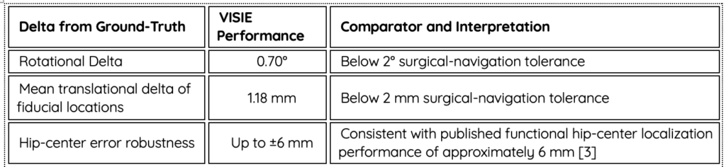
Table 1. Summary of the anatomic-registration accuracy relative to ground truth and the tested robustness to hip-center localization error.
Accurate femoral registration is foundational to successful computer-assisted TKA, yet conventional registration workflows can be limited by the need for invasive fixation, extensive anatomic exposure, and manual point-by-point registration. As surgical approaches continue to evolve toward tissue-sparing techniques, navigation technology must adapt to the limited anatomic visibility encountered in modern procedures.
The VISIE registration approach addresses this challenge by enabling rapid, pinless anatomic registration using a high-quality three-dimensional optical acquisition of the exposed femur. By leveraging advanced optical scanning and anatomy-based registration, the system is designed to establish accurate alignment between intraoperative anatomy and preoperative CT models, even when only a limited portion of the femur is visible.
The technology was first demonstrated under extreme visibility constraints using a porcine knee model, where successful alignment was achieved despite intentionally minimizing the available femoral surface. This evaluation provided qualitative evidence of the robustness of the registration approach under challenging anatomic conditions.
The approach was further evaluated in a human cadaveric TKA model using a subvastus surgical approach, representing a clinically relevant and highly constrained exposure. Despite limited visualization of the distal femur and the inherent differences between CT-derived bony anatomy and optical scans capturing cartilage and soft tissue surfaces, the registration achieved a rotational difference of 0.70° and a mean translational difference of 1.18 mm relative to an independently established fiducial-based reference. These results demonstrate accuracy within commonly accepted navigation performance targets.
[1] Umeyama S. IEEE Trans Pattern Anal Mach Intell. 1991;13(4):376–380. doi:10.1109/34.88573.
[2] Li P, Wang R, Wang Y, Tao W. IEEE Access. 2020;8:68030–68048. doi:10.1109/ACCESS.2020.2986470.
[3] Speirs AD, Benoit DL, Beaulieu ML, Lamontagne M, Beaulé PE. HSS J. 2012;8(3):192–197. doi:10.1007/s11420-012-9296-6.